












 中国 – 中文

糖尿病患者种植修复与种植体的合理选择
针对糖尿病患者普遍存在的咀嚼功能受限、缺牙率较高的现状(我国糖尿病患病人数已达1.18亿),临床中常遇患者因担心种植失败而将种植修复视为“禁区”。
然而,现有循证医学证据表明:糖尿病患者并非种植治疗的绝对禁忌人群。关键在于全面评估并控制血糖及相关影响因素,同时选择具备先进表面处理技术、良好骨结合性能的种植体系统。在严格把握适应证,科学开展种植修复可使糖尿病患者重获稳定、有效的咀嚼功能。
糖尿病对种植修复的三类主要临床影响
糖尿病患者在种植治疗过程中面临的挑战显著高于普通人群,其核心机制均与高血糖状态密切相关,具体表现为以下三个方面:
影响一:骨结合质量下降,骨代谢失衡加剧
牙槽骨是种植体获得初期稳定性和远期存留率的基础。糖尿病可加速牙槽骨的吸收进程,导致骨量减少、骨密度降低,即便同期实施引导骨再生术,其成骨效果也可能受限。
同时,高血糖还会破坏身体里成骨细胞和破骨细胞的平衡,延缓种植体与颌骨的“骨结合”过程。骨结合效率的下降将影响种植体的初期稳定性,进而增加早期失败风险。
影响二:术后愈合延迟,感染风险升高
高血糖可抑制免疫细胞(如中性粒细胞、巨噬细胞)的趋化与吞噬功能,导致术后局部防御能力下降。临床观察显示,糖尿病患者的种植术后创面愈合缓慢、新生骨质不佳或者骨量不足【24】。术后种植体周炎的发生风险比普通人高50%【25】。
此外,糖尿病常合并微血管病变,引起口腔局部血供减少,组织修复所需的氧气及营养供应不足,进一步加重愈合延迟。
影响三:长期维护难度大,种植体周围炎发病率升高
高糖尿病与牙周炎之间存在双向促进关系:血糖控制不佳会加重牙周组织炎症,而持续的牙周炎症又反作用于全身糖代谢,形成恶性循环。
因此,糖尿病患者术后发生种植体周围炎的风险显著高于健康人群。若长期血糖控制不理想,牙槽骨吸收速度加快,最终可导致种植体周围支持结构丧失,出现种植体松动甚至脱落,严重影响种植体的远期存留率。
糖尿病患者种植体选择
鉴于糖尿病对骨结合、创面愈合及长期抗感染能力带来的多重影响,种植体的合理选择成为决定治疗成败的关键环节。并非所有种植体系统均适用于糖尿病患者,临床中应重点关注以下三项核心性能指标:
1:高效的骨结合能力
优先选择具备促进骨结合进程的表面处理技术(如活性亲水性表面),以缩短骨结合周期,增强成骨细胞黏附与增殖能力。这对于自身骨结合能力较弱的糖尿病患者尤为重要,有助于提高种植体初期稳定性及远期存活率。
2:良好的生物相容性
糖尿病患者常处于慢性低度炎症状态,对植入材料的免疫反应较为敏感。应选择生物相容性优异、致敏性低的种植体材质与表面涂层,以降低异物反应及术后感染风险。
3:足够的机械强度与结构设计
由于糖尿病患者常伴有骨量不足或骨密度下降问题,种植体需具备较高的抗疲劳强度及合理的螺纹形态设计,以提供足够的支撑力和长期稳定性,避免因骨质量差而出现的力学失败。
临床策略:钛锆亲水种植体在糖尿病患者种植修复中的应用
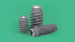
采用高强度钛锆合金材质,结合活性亲水表面的种植体已成为具备循证依据的临床策略。该方案通过促进早期成骨、调节炎症环境及加速愈合进程,提升糖尿病患者的种植成功率。
亲水表面处理技术—加速骨结合进程
活性亲水表面技术自2005年问世以来,已被证实可显著改善种植体骨结合效率。其核心机制在于增强种植体表面的血液亲和性。通过特殊工艺在喷砂酸蚀形成的粗糙表面基础上实现超亲水特性(接触角为0°)【1】,可在植入后10分钟内快速吸附血液中的蛋白质【2,3】,提速成骨早期血管化的发生【4,5,6】。
相较于未做亲水处理的表面,亲水表面技术使得种植体表面产生纳米结构,可让种植体与骨组织的有效接触面积提升50%【22-23】,并将原本需要数月的骨结合周期缩短至3–4周【11-14】。对于伤口愈合显著延迟的糖尿病患者而言,这一技术可有效降低愈合期内的感染及早期失败风险,使种植体更快、更稳定地实现骨整合。

钛锆合金材质应对骨量不足的力学保障
针对糖尿病患者牙槽骨量不足、骨密度低的问题,钛锆合金相较于纯钛具有更高的机械强度和更好的生物相容性【15-16】。对于骨条件欠佳的糖尿病患者,钛锆合金材质可在不牺牲生物学性能的前提下提供更强的初期稳定性与长期负载能力。

抗炎特性与20年长期临床验证
亲水表面处理与钛锆合金材质的协同应用,不仅提升了骨结合效率与力学性能,同时赋予了种植体更强的抗炎能力。其机制在于通过免疫调节作用,抑制炎症相关因子的过度释放,从根本上降低糖尿病患者种植体周围炎的发生风险。
在2017年第95届国际牙科研究会(IADR)上形成的专家共识指出:亲水表面可有效调节由糖尿病诱导的炎性反应,改善骨结合【17-18】。

该方案的有效性已获得多项临床研究的支持。现有数据显示,糖尿病患者接受钛锆合金联合亲水表面处理种植体修复后,2年种植体存活率可达100%【19-20】;术后5年随访亦证实种植体周围骨水平能够维持稳定【21】。上述结果表明,为糖尿病患者科学选择适宜的种植体系统,不仅有助于实现短期内的种植成功,更可保障种植体的远期稳定与功能持久。
恢复咀嚼功能,始于科学种植
血糖的有效控制及种植体的科学选择是糖尿病患者种植治疗成功的关键因素。
针对高血糖所致的愈合延迟、骨结合障碍及感染风险,采用亲水表面处理的钛锆合金种植体已展现出充分的临床循证支持,为该人群提供了可预期的修复方案。
参考文献
1. Rupp F et al. : Enhancing surface free energy and hydrophilicity through chemical modification of microstructured titanium implant surfaces. Journal of Biomedical Materials Research A, 76(2):323-334, 2006.
2. DeWild M : Superhydrophilic SLActive® implants. Straumann document 151.52, 2005.
3. Maniura K : Laboratory for Materials – Biology Interactions Empa, St. Gallen, Switzerland Protein and blood adsorption on Ti and TiZr implants as a model for osseointegration. EAO 22nd Annual Scientific Meeting, October 17 – 19 2013, Dublin.
4. Schwarz F et al. : Bone regeneration in dehiscence-type defects at non-submerged and submerged chemically modified (SLActive®) and conventional SLA® titanium implants: an immunohistochemical study in dogs. J Clin.Periodontol. 35.1 (2008): 64– 75.
5. Rausch-fan X et al. : Differentiation and cytokine synthesis of human alveolar osteoblasts compared to osteoblast-like cells (MG63) in response to titanium surfaces. Dental Materials 2008 Jan;24(1):102-10. Epub 2007 Apr 27.
6. Schwarz F et al. : Histological and immunohistochemical analysis of initial and early osseous integration at chemically modified and conventional SLA® titanium mplants: Preliminary results of a pilot study in dogs. Clinical Oral Implants Research, 11(4): 481-488, 2007.
7. Lang, NP et al. : Early osseointegration to hydrophilic and hydrophobic implant surfaces in humans. Clin Oral Implants.Res 22.4 (2011): 349–56.
8. Raghavendra S et al.: Early wound healing around endosseous implants: a review of the literature. Int. J. Oral Maxillofac. Implants. 2005 May–Jun;20(3):425–31.
9. Oates TW et al. : Enhanced implant stability with a chemically modified SLA® surface: a randomized pilot study. Int. J. Oral Maxillofac. Implants. 2007;22(5):755–760.
10. Schwarz F et al. : Bone regeneration in dehiscence-type defects at chemically modified (SLActive) and conventional SLA titanium implants: A pilot study in dogs. J. Clin. Periodontol. 2007;34(1):78–86.
11. Buser D et al. : Enhanced bone apposition to a chemically modified SLA titanium surface. J. Dent. Res. 2004 Jul;83(7):529–33.
12. Schwarz F et al. : Histological and immunohistochemical analysis of initial and early subepithelial connective tissue attachment at chemically modified and conventional SLA® titanium implants. A pilot study in dogs. Clin. Oral Impl. Res. 2007;11(3):245–455.
13. Schwarz F et al. : Effects of surface hydrophilicity and microtopography on early stages of soft and hard tissue integration at non-submerged titanium implants: An immunohistochemical study in dogs. J. Periodontol. 2007;78(11):2171–2184.
14. Zöllner et al. : Immediate and early non-occlusal loading of Straumann implants with a chemically modified surface (SLActive®) in the posterior mandible and maxilla: interim results from a prospective multicentre randomized-controlled study. Clinical Oral Implants Research, 19(5), 442-450,2008.
15. Bernhard N, Berner S,de Wild M, et al. The Binary TiZr Alloy – A Newly Developed Ti Alloy for Use in Dental Implants. Forum Implantologicum 2009 5(1) 30-39
16. Straumann, data on file
17. Hotchkiss K.M, Sowers K.T, Olivares-Navarrete R. Clinical Implants Differentially Modulate Inflammatory Response and Osteogenic Differentiation. Presented at 95th General Session of International Association for Dental Research, USA, March 22-25, 2017. https://iadr2017.zerista.com/event/member?item_id=5725415
18. Lee R, Hamlet SM, Ivanovski S. The influence of titanium surface characteristics on macrophage phenotype polarization during osseous healing in type I diabetic rats: A pilot study. Clin Oral Impl Res
19. Cabrera-Domínguez, José J et al. “Clinical performance of titanium-zirconium implants with a hydrophilic surface in patients with controlled type 2 diabetes mellitus: 2-year results from a prospective case-control clinical study.” Clinical oral investigations vol. 24,7 (2020): 2477-2486
20. Friedmann, Anton et al. “One-year performance of posterior narrow diameter implants in hyperglycemic and normo-glycemic patients-a pilot study.” Clinical oral investigations vol. 25,12 (2021): 6707-6715
21. Haimov, Eliezer et al. “Differences in Titanium, Titanium-Zirconium, Zirconia Implants Treatment Outcomes: a Systematic Literature Review and Meta-Analysis.” Journal of oral & maxillofacial research vol. 14,3 e1. 30 Sep. 2023
22. Wennerberg A, Galli S, Albrektsson T, et al. Current knowledge about the hydrophilic and nanostructured SLActive surface. Clin Cosmet Investig Dent. 2011 Sep 5:3:59-67
23. Straumann, Developed area ratio by nanostructures on Rxd modMA surface. Report SR0748 2017
24. Zhang S ,Song S ,Wang S ,et al. Type 2 diabetes affects postextraction socket healing and influences first-stage implant surgery: a study based on clinical and animal evidence[J]. Clin Implant Dent Relat Res, 2019,21(3):436-445.
25. Monje A ,Catena A ,Borgnakke WS . Association between diabetes mellitus/hyperglycaemia and peri-implant diseases: systematic review and meta-analysis[J]. J Clin Periodontol, 2017,44(6):636-648.
*本文章仅供医学专业人士参考,具体需结合产品说明书及临床实际情况予以判断。